-Trusted by 100+ veterinary clinics. - Completed 300,000+ diagnostic tests for farmers. - Accredited by VETQAS.

This guide is educational only. Always consult with your own veterinarian.

Your vet has experience and knowledge on your land type and parasite infection patterns.

GutWorm Faecal Egg Counts (FEC)

FEC testing gives you a guide of the level of parasitic worms inside your animals. In cattle and sheep there are approximately 20 species of gutworms which may cause a range of diseases when in numbers. In animals, the parasitic gutorms types include roundworms, whipworms, hookworms and tapeworms. These parasites live inside the animal and usually cause organ damage as well as disrupting animals ability to absorb nutrients. Low level of gutworm infection in animals can be beneficial as it allows the animal to develop its own immunity against the parasites. 

Gutworms may cause major disease in animals, and cause economic losses for the farmer, such as; 

  • Failure to thrive
  • Anemia
  • Poor health
  • Weight loss
  • Diarrhea
  • Death- In some cases.

The gutworms which cause the most illness in cattle are Cooperia and Ostertagia. In sheep the most significant are Haemonchus, Nematodirus, Teladorsagia and Trichostrongylus. In horses, the most common are Cyathostominae (blood worm), Strongylus and Parascaris.

It takes at least 3 weeks for the gutworm parasites to develop inside the animal and start laying eggs. This means the animal must be on grass for 3+ weeks before the worm eggs can be detected in the lab. When animals are turned out in spring to graze, gutworm larvae can be present on the grass as they can survive the winter's harsh conditions. These larvae can then infect the animals when suitable weather conditions occur. Most animals develop immunity to gutworms over their lifetimes but other diseases can tax the immune system at any age and allow the parasites to reinfect. Exposure to gutworm parasites can be reduced by grazing management, cross-species grazing and reducing stocking rates. 

The results from the Gutworm Faecal Egg Counts (FEC) inform you on parasite types detected and estimate eggs per gram (EPG) present. There is a known correlation between eggs per gram and the number of parasitic adult worms present in the animal gut & intestines. The results of this test will tell you the count and the type of worms, such as the Trichostrongyle family in cattle and sheep. To distinguish between the different Trichostrongyle eggs a larval hatch may be carried out, but this is infrequently required. Gutworm testing supplies detailed information so you can make decisions on drug treatment, grazing patterns and administration plans. This test can also detect resistance or if the treatment worked by a post-dosing faecal egg count/drench test (FEC test) or a faecal egg count reduction test (FECRT).

Resistance of anthelmintics and flukicides (parasite drugs) is now a large issue. Anthelmintic/flukicide resistance occurs when worms/fluke survive a dose of a wormer/fluke treatment that would normally be expected to kill them. Faecal Egg Count (FEC) & Sedimentation is vital to monitor anthelmintic/drugs effectiveness. Many farms in Ireland already have some form of anthelmintic (treatment) resistance present in their livestock, which may cause major production & financial loss.

Liver Fluke

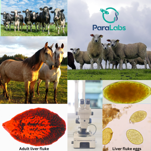
Liver fluke (Fasciola hepatica) is a flatworm parasite, the adults are the size of a €2 coin. Fasciolosis (liver fluke infestation) is one of the major causes of parasitic disease in cattle, sheep and horses. 

Liver fluke causes major disease in animals, many of which cause financial losses for the farmer,  such as; 

  • Low milk production
  • Decreased fertility
  • Failure to thrive
  • Weight loss
  • Longer finishing time
  • Condemned livers at slaughter

Due to Ireland's wet climate, the prevalence of liver fluke is high on farms as these are the ideal conditions for its intermediate host, the mud snail (Galba truncatula). In particular, damp and marshy areas with poor drainage are perfect conditions for this snail. Once the animal grazes on grass infested with this larvae, the parasite takes 12 weeks to reach adulthood where it will begin laying eggs.  This means that young animals must be grazing on grass for a minimum of 3 months before checking for liver fluke infections. Liver fluke infections are hard to visually diagnose, but chronic cases result in bottle jaw, jaundice and ill thrift.

The sedimentation technique is used to identify the fluke eggs present in the faeces. Both rumen and liver fluke eggs are detected in this technique, so,  differentiating between rumen and liver fluke is very important as these cause 2 different infections. 

A negative sedimentation results does not mean that there is no infection, there are different reasons such as; 

  • Early stage infections - the larvae have not matured to adulthood, therefore, they do not produce eggs. However, early stages of liver fluke can still cause substantial liver damage.
  • Intermittent shedding of eggs - Animals do not constantly shed liver fluke eggs, meaning that an infected animal may have no liver fluke eggs in its faeces at the time of testing, even when infected. 

If you receive a negative result, but liver fluke is still suspected, retest in 6 weeks to rule out infection. Unlike gutworms, there is no evidence that animals develop immunity to liver fluke parasites, so they have to be detected and controlled by testing and flukicide treatment.


banner_image

Rumen Fluke

Rumen/Stomach fluke is a parasite that infects livestock such as cattle, sheep and goats (ruminates). The lifecycle of rumen fluke is similar to that of liver fluke, involving a snail as an intermediate host. The adult stomach fluke lives in the animal's rumen, is shaped like a broad bean and can cause health issues in heavily infected animals. The prevalence of rumen fluke in livestock is higher in regions with wet and heavy ground. 


Stomach fluke infestations may lead to notable economic losses such as;  

-Decreased milk yield 

-Reduced weight gain 

-Ill thrift - Dehydration, Diarrhoea 

-Mortality (in some cases) high immature presence

Immature stomach fluke infections can be particularly severe in young cattle and sheep. These infections occur when the immature flukes, which attach themselves in the upper small intestine, cause lesions, blood loss and significant damage to the intestinal lining. This can lead to symptoms such as severe diarrhea, dehydration, anorexia, and unthriftiness. In heavy infestations, the damage can be extensive, leading to high mortality rates. 

Detection of stomach fluke eggs in faeces is typically done using sedimentation technique. Identifying rumen fluke eggs involves distinguishing it from liver fluke through microscopic inspection. Fluke eggs usually become detectable 8-10 weeks (about 3 months) after infection, requiring routine monitoring of animals that graze in high-risk areas

banner_image

Lung Worm

The lungworm parasite is similar to gutworm but they mainly reside in the lungs of an animal. The parasites infect the lower respiratory tract where clinical signs of infection include coughing and difficulty in breathing. The lungworm infection may lead to secondary infections which result in pneumonia or bronchitis.  

Lungworm infection causes a serious and often fatal disease in animals, which may result in financial losses for the farmer, such as; 

·      Failure to thrive

·      Irreversible lung damage.

·      Poor health.

·      Low milk yield

·      Susceptible to other respiratory pathogens

·      Death- In some cases. 

In Ireland the lungworm species infecting cattle is Dictyocaulus viviparus. In sheep & goats the most common species are D. filaria, P. rufescens and M. capillaris. In horses and donkeys the lungworm is D arnfieldi. Other lungworm species occur but are not common. Lungworm is host specific, meaning the lungworm of one animal species will not infect another species. (e.g: Bovine lungworm will not infect ovine.)

The lungworm life cycle takes about 2-4 weeks where they reproduce by producing eggs in the lungs. The eggs are coughed up and swallowed where they hatch into larvae in the digestive system and pass through the animal into the faeces. When the larvae are in the dung pat they develop further and spread around the field. The larvae on the grass are then eaten by the animal and burrow through the small intestine. They mature and travel through the lymph and blood system to the lungs where they cause disease. 

Lungworm mainly affects younger animals with no previous exposure to the parasite. There is a rising occurrence of adult animals getting lungworm infections. This happens when heavy infections overcome the immune system, even when there was pre-existing immunity present. Also it happens when adult animals that previously never came in contact with lungworms can easily become infected. 

For lungworm to be diagnosed in the lab, the animal must be actively coughing. When you see them coughing, the animal is removing the lungworm eggs from the lungs and passing larvae in the faeces. In the lab we perform a modified Baermann technique where the larvae migrate from the faeces into a suspended liquid. The contents are then microscopically examined where the larvae are measured and diagnosed. It is very important that the sample for this test is taken directly (rectally) from the animal. This is because in the environment there are free living larvae which contaminate the sample and make lungworm larvae less detectable.

The three major groups of treatments (white/yellow/clear) are all effective against lungworm. Each drug group kills the lungworm in different time frames. It is highly advisable to get veterinary advice on which product before starting treatment to avoid secondary infection (pneumonia).

Shopping Cart

    Your cart is empty